Condition Focus
Cartilage Preservation
Surgical and biologic strategies to protect the remaining joint surface, address chondral defects, and delay or prevent hip arthritis.
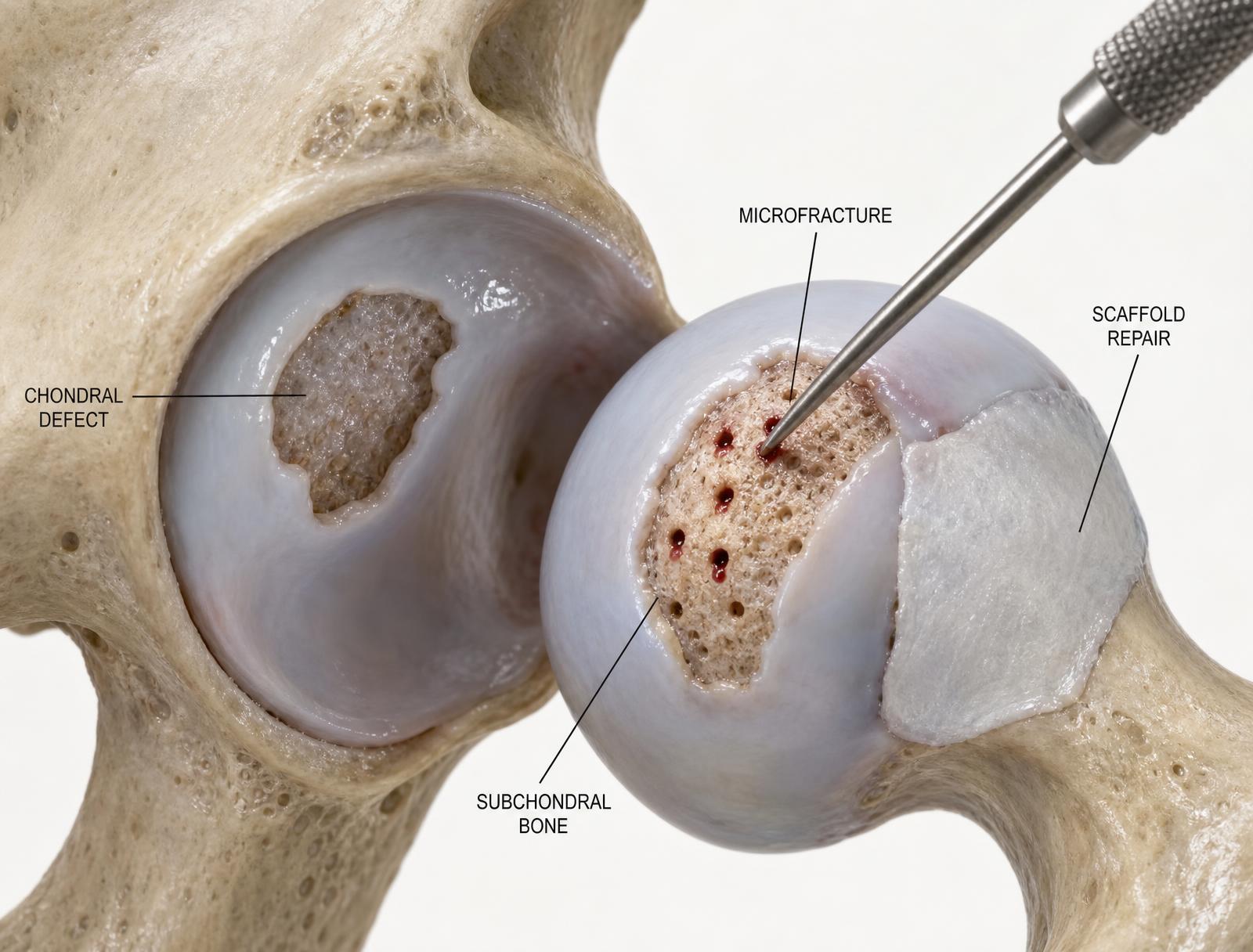
Articular cartilage in the hip has almost no capacity to heal itself. Once a chondral flap or delamination develops — usually driven by cam impingement, dysplastic rim overload, or trauma — the defect tends to enlarge unless the mechanical cause is corrected.
Cartilage preservation therefore has two parts: fixing the mechanics that damaged the surface, and treating the defect itself with debridement, marrow stimulation, or a scaffold-based technique matched to the lesion's size and location.
Symptoms & findings
- /Deep, aching hip pain that persists after activity
- /Pain with weight-bearing rotation rather than a discrete catching sensation
- /Stiffness and loss of terminal rotation
- /Swelling or a heavy feeling after prolonged loading
- /Progressive symptoms despite prior arthroscopy or therapy
Diagnostic approach
Weight-bearing radiographs assess joint space and the bony morphology loading the surface. MRI — ideally with cartilage-sensitive sequences — grades chondral injury and detects subchondral edema and delamination. Lesion size, containment, and remaining joint space determine whether preservation or arthroplasty is appropriate.
Treatment options
Correct the mechanical cause
Cam and pincer osteoplasty, labral repair to restore the fluid seal, or acetabular reorientation for dysplasia. Treating a chondral defect without correcting the load that created it leads to recurrence.
Debridement and microfracture
Unstable cartilage flaps are debrided to a stable rim, and small contained defects are treated with marrow stimulation to recruit a fibrocartilage repair tissue.
Scaffold-based repair
For larger or delaminated lesions, a scaffold or biologic augment is used to support a more organized repair tissue and improve durability compared with microfracture alone.
Load management and rehabilitation
Protected weight bearing, controlled motion, and a graded return to loading are essential to protect immature repair tissue during the first several months.
Why patients see Dr. Almasri in Cincinnati
- /Dual fellowship training in hip preservation surgery and orthopedic sports medicine
- /Practice strongly focused on the young and active hip
- /Team physician experience with FC Cincinnati and regional collegiate programs
- /Peer-reviewed research and international faculty roles in hip preservation
- /In-office and direct online scheduling through Mercy Health