Condition Focus
Hip Arthroscopy
Minimally invasive access to the hip joint through two to three small portals — for labral, cartilage, ligamentum teres, and capsular pathology.
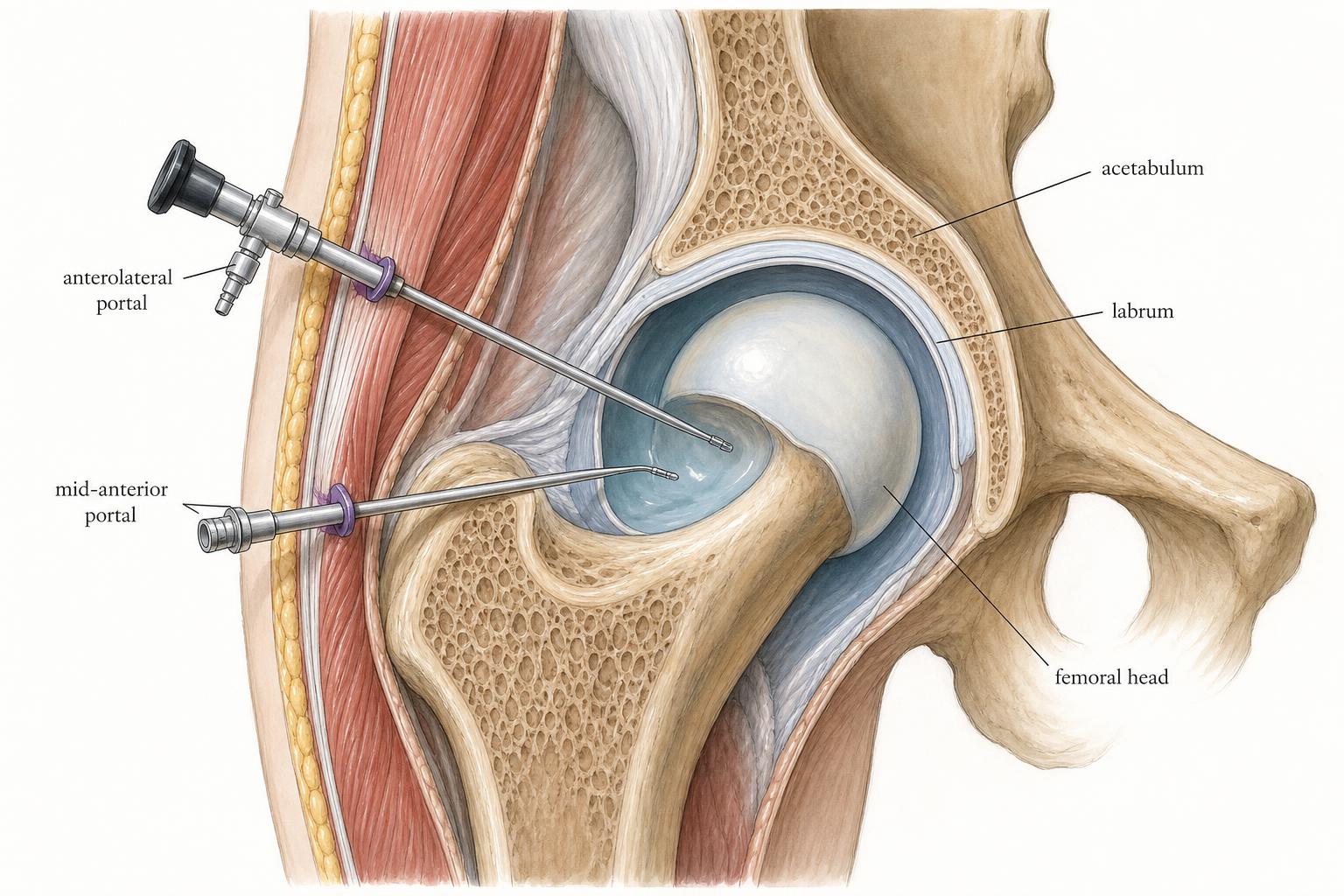
Hip arthroscopy allows direct treatment of intra-articular pathology through portals a few millimeters wide. A camera and long instruments reach both the central compartment — labrum, cartilage, ligamentum teres — and the peripheral compartment, where cam bone is reshaped.
The technical detail that matters most is what happens at the end of the case: the capsule is closed to preserve the hip's soft-tissue restraints. Skipping closure is a recognized cause of post-arthroscopy instability and revision surgery.
Symptoms & findings
- /Mechanical symptoms: catching, clicking, or locking inside the joint
- /Deep groin pain reproduced by flexion and internal rotation
- /Pain unresponsive to a well-executed course of physical therapy
- /Imaging showing a labral tear, cam or pincer morphology, or a chondral flap
- /Positive response to a diagnostic intra-articular injection
Diagnostic approach
Candidacy is determined by matching symptoms and examination to imaging findings. Radiographs quantify cam and pincer morphology and screen for joint space narrowing; MRI defines labral and chondral injury. Patients with advanced arthritis or significant uncorrected dysplasia are generally better served by other procedures.
Treatment options
What can be treated arthroscopically
Labral repair and reconstruction, cam and pincer osteoplasty, chondral debridement and microfracture, ligamentum teres debridement, synovectomy, loose body removal, capsular plication and closure, and endoscopic treatment outside the joint such as iliopsoas and abductor pathology.
The procedure
Under anesthesia, the hip is accessed through two to three portals. Traction is used only as long as necessary for central compartment work to protect the nerves and cartilage, and the case is completed in the peripheral compartment with the hip flexed. The capsule is repaired before closure.
Recovery
Most patients go home the same day on crutches with a defined range-of-motion protocol. Physical therapy starts within days, crutches come off around 2–6 weeks depending on what was performed, and return to sport is typically 4–6 months.
Why patients see Dr. Almasri in Cincinnati
- /Dual fellowship training in hip preservation surgery and orthopedic sports medicine
- /Practice strongly focused on the young and active hip
- /Team physician experience with FC Cincinnati and regional collegiate programs
- /Peer-reviewed research and international faculty roles in hip preservation
- /In-office and direct online scheduling through Mercy Health